Bodyweight Squat
The fundamental lower-body pattern for developing functional strength, mobility, and kinetic awareness.
Biomechanics Analysis
Closed Kinetic Chain Dynamics
The bodyweight squat operates as a closed kinetic chain movement, meaning the distal segment (the feet) remains fixed to the ground. Movement relies on Triple Extension, where the Gluteus Maximus (hip), Quadriceps (knee), and Soleus (ankle) contract concentrically to drive the Center of Mass (COM) upward against gravity. The Ground Reaction Force (GRF) is transferred through the kinetic chain, necessitating synchronized joint articulation.
Torque & Moment Arms
Primary torque (τ) is generated at the acetabulofemoral (hip) and tibiofemoral (knee) axes. The magnitude of torque at each joint depends on the horizontal distance (moment arm) between the joint axis and the line of force (gravity acting on the COM). Proper mechanics require the tibia and torso to maintain a roughly parallel alignment at the bottom of the movement to distribute load evenly between the anterior and posterior chains.
Stabilization & Intra-Abdominal Pressure
Stabilization is not passive; it requires active isometric engagement of the Transversus Abdominis and Erector Spinae. This creates intra-abdominal pressure (IAP), neutralizing spinal shear forces and maintaining a neutral lumbar spine (lumbar flexion θ ≈ 0°). This rigidity transfers force efficiently from the legs to the torso.
Muscle Activation Map
Primary Movers (Agonists)
- ▸Quadriceps Femoris: Knee extension (Rectus Femoris, Vastus Lateralis/Medialis/Intermedius).
- ▸Gluteus Maximus: Hip extension and external rotation.
Synergists
- ▸Adductor Magnus: Assists in hip extension.
- ▸Soleus: Plantarflexion to stabilize the tibia.
- ▸Hamstrings: Dynamic knee stabilization (co-contraction).
Deep Stabilizers
- ▸Erector Spinae: Maintains spinal extension.
- ▸Transversus Abdominis: Compresses abdominal viscera (IAP).
- ▸Multifidus: Segmental spinal stability.
Execution Protocol
The Setup
Stand with feet slightly wider than shoulder-width apart. Rotate toes outward approximately 15°–30° to align with the natural tracking of the femur. Engage the core (bracing) and keep the chest proud.
The Descent (Eccentric Phase)
Simultaneously unlock the hips and knees. Think about "sitting back and down" between your heels. Ensure the knees track directly over the second and third toes to prevent valgus collapse. Maintain a neutral spine; do not allow the lumbar to round.
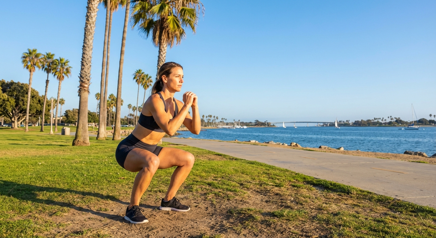
The Bottom Position
Descend until the hip crease is at or below the top of the knee (parallel depth), provided mobility allows without spinal compensation. Maintain tension; do not bounce passively off the joints.
The Ascent (Concentric Phase)
Drive through the mid-foot (creating a tripod foot with the heel, big toe knuckle, and little toe knuckle). Exhale forcefully as you extend the hips and knees simultaneously, returning to the standing position with full glute contraction.
Common Faults & Corrections
| Fault | Biomechanics Issue | Correction |
|---|---|---|
| Knee Valgus (Caving In) | Weak external rotators (Glute Medius) or poor adductor flexibility. Increases ACL stress. | Cue "spread the floor apart" with your feet; drive knees outward over toes. |
| Butt Wink | Posterior pelvic tilt at the bottom (lumbar flexion > 0°). Often due to anatomy or tight hamstrings. | Limit depth to the point just before the pelvis tucks. Improve ankle mobility. |
| Heels Lifting | Limited dorsiflexion range of motion at the talocrural joint. | Widen stance slightly or perform ankle mobility drills. Keep weight over the mid-foot. |
| Thoracic Collapse | Loss of thoracic extension, shifting center of mass forward (moment arm increases at lumbar). | Keep eyes on the horizon. Cue "show the logo on your shirt" to the wall in front. |